41 diagram of villus
The diagram shows the structure of villus in the small intestine. The arrows show the direction of flow of fluids. After a meal of fried chicken, where will minute fat globules be present in the largest amount? A. Part A. B. Part B. C. Part C. D. Part D. 11. Lipase solution was added to milk. After 30 minutes, the milk had become more acidic. Sep 20, 2018 - villi diagram simple kidney diagram bladder diagram parts of a villus small intestine crypts.
Oct 26, 2021 · Fig. 1: Schematic diagram of paracrine Hh signalling in the mammalian intestine. ... and are highly expressed in the endoderm of the intestine before villus formation in the embryo .

Diagram of villus
The study aimed to explore the effects of fortified fermented rice-acid on the antioxidant capacity of mouse serum and the gut microbiota. Hair characteristics, body mass index, intestinal villus height, intestinal crypt depth, serum antioxidant capacity, and gut microbiota of mice were first measured and the correlation between the antioxidant capacity of mouse serum and the gut microbiota ... 1580s, "to make a map or diagram of, lay down on paper according to scale;" also "to lay plans for, conspire to effect or bring about" (usually with evil intent), from plot (n.). Intransitive sense of "to form a plan or device" is from c. 1600. Related: Plotted; plotter; plotting. The picture above is a diagram of what is inside the villus. It explains what kind of nutrients is absorbed by the blood capillary which is glucose, ...
Diagram of villus. "light, feathery stuff," 1790, apparently a variant of floow "wooly substance, down, nap" (1580s), perhaps from Flemish vluwe, from French velu "shaggy, hairy," from Latin vellus "fleece," or Latin villus "tuft of hair" (see velvet). OED suggests fluff as "an imitative modification" of floow, "imitating the action of puffing away some light substance." Slang bit of fluff "young woman" is from 1903. The marshmallow confection Fluff dates to c. 1920 in Massachusetts, U.S. Intestinal villi (singular: villus) are tiny, finger-like projections that protrude from the epithelial lining of the intestinal wall.2 pages An alternate mode of infection consists of internal autoinfection, where the eggs release their hexacanth embryo, which penetrates the villus continuing the infective cycle without passage through the external environment . The life span of adult worms is 4 to 6 weeks, but internal autoinfection allows the infection to persist for years. Chromosome 21 is one of the 23 pairs of chromosomes in humans.Chromosome 21 is both the smallest human autosome and chromosome, with 48 million base pairs (the building material of DNA) representing about 1.5 percent of the total DNA in cells.Most people have two copies of chromosome 21, while those with three copies of chromosome 21 have Down syndrome, also called "trisomy 21".
Syncytiotrophoblast (from the Greek 'syn'- "together"; 'cytio'- "of cells"; 'tropho'- "nutrition"; 'blast'- "bud") is the epithelial covering of the highly vascular embryonic placental villi, which invades the wall of the uterus to establish nutrient circulation between the embryo and the mother. It is a multi-nucleate, terminally differentiated syncytium, extending to 13 cm. The diagram shows the structure of a villus. In the small intestine, some of the products of digestion are absorbed into the blood by active transport. (a) Explain what is meant by active transport. _____ _____ Villi increase the internal surface area of the intestinal walls making ... villus diagram | science biology diagrams | drawing for students ... Sep 11, 2017 ... The transfer of food particles from the digestive system to the circulatory system takes place at the inner lining of the small intestine, ...
1670s as a technical term in perspective drawing; more generally by 1706 as "the representation of anything drawn on a plane; a drawing, sketch, or diagram of any object," from French plan "ground plot of a building, map," literally "plane surface" (mid-16c.), from Latin planum "level or flat surface," noun use of adjective planus "level, flat" (from PIE root *pele- (2) "flat; to spread"). The notion is of "a drawing on a flat surface." A doublet of plain via a later, learned French form. The meaning "scheme of action, formulated scheme for the accomplishment of some object or attainment of an end" is by 1713. Aug 13, 2020 — This is a diagram of an intestinal villus. The thin surface layer is drawn above. Intestinal villus: An image of a simplified structure of ... For example, human chorionic villus samples, bone marrow, etc. In case these samples are difficult to find, lymphocytes can be taken and induced to divide. Q.4. What happens if a karyotype test is abnormal? Ans: Abnormal karyotype results may indicate the presence of a genetic anomaly in an individual. Q.5. a, Phase diagram of crypt morphologies (\(\tilde \kappa _0 = 6\), φ = 0.2) as a function of normalized crypt apical tension m and villus cell volume v ev, showing the villus cell swelling favors ...
Image from page 713 of "Kirkes' handbook of physiology" (1907)
early 14c., probably from Old Provençal veluet, from Vulgar Latin *villutittus, diminutive of Vulgar Latin *villutus "velvet," literally "shaggy cloth," from Latin villus "shaggy hair, nap of cloth, tuft of hair," probably a dialectal variant of vellus "fleece," from PIE *wel-no-, suffixed form of *uelh- "to strike" (see svelte).

Image from page 275 of "Embryology" (1949)
1706, also velure, velours, from French velours "velvet," from Old French velor, alteration of velos "velvet," from Old Provençal velos, from Latin villosus (adj.) "shaggy, hairy, rough" (in Medieval Latin "velvet"), from villus "shaggy hair, tuft of hair" (see velvet).
Digestive System | Basicmedical Key
The figure below is a diagram of an intestinal villus. Study it and answer the questions that follow. Name the parts labeled A - D (2 mks) What is the importance of the villi? (1 mk) What is the function of the part labeled F (1 mk) Most of absorption of digested food in mammals takes place in the ileum.

Image from page 116 of "A treatise on the science and practice of midwifery" (1878)
The circle of Willis is located on the inferior surface of the brain within the interpeduncular cistern of the subarachnoid space encircles various structures within the interpeduncular fossa (depression at the base of the brain) including the optic chiasm and infundibulum of the pituitary gland.. Although significant anatomic variations exist, the circle of Willis is typically composed of ...

Image from page 700 of "Elements of human physiology" (1907)
"long, slender hair," 1704, plural villi, from modern use of Latin villus "tuft of hair, shaggy hair, wool, fleece" (see velvet).

Image from page 60 of "Animal biology" (1940)
"observation or diagram of the heavens, showing the positions of planets, on any given day, used by astrologers," mid-16c., from French horoscope, from Latin horoscopum/horoscopus, from Greek hōroskopos "nativity, horoscope," also "one who casts a horoscope, one who observes the hour of a birth," from hōra "hour; season; period of time" (see hour) + skopos "watcher; what is watched" (from PIE root *spek- "to observe"). The notion is of "observing the hour" (of someone's birth, etc.). The word was in late Old English and Middle English as horoscopum, from Latin, but the modern form is considered to be a reborrowing. Related: Horoscopic; horiscopal. Horoscopy "the casting of a nativity" is attested from 1650s, from Latin horoscopium, from Greek hōroskopeion, from hōroskopia.

Image from page 275 of "Animal biology; Human biology. Parts II & III of First course in biology" (1910)
g Schematic diagram of E. tenella lifecycle, source: ... Budding enteroids can be developed from villus tips in the embryonic chicken because almost all villus cells in poultry are proliferative at hatch with cell mitosis playing an important role in post-hatch hyperplasia 27. The enteroid transcriptome indicated the embryonic cultures continue ...
Trophoblast - wikidoc
This villus consists of the blood capillaries. These blood capillaries absorb the digested food from the small intestine. It is long and more coiling as compared to the large intestine. But it has a small diameter. a) Duodenum is the first part of the small intestine. This part connects to the liver and pancreases.

Image from page 884 of "Anatomy, descriptive and surgical" (1887)
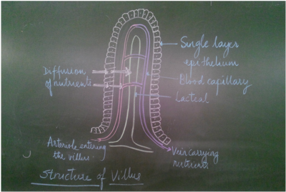
These answers refer to the questions and diagrams on the slides of this Short presentation of villus structureHow is a villus structure related to its role in absorption and transport of the products of digestion ?Describe the shape. It is a tall thin finger shaped structure.What is inside a villus? It has a lacteal, blood capillaries, and small arterioles or veins What are the surface cells ...

Chrysis sp., Brookside Gardens, Mont Co, 5-15-16
"pertaining to schemes," 1701, from Latin stem of scheme (n.) + -ic. Noun meaning "diagram" is first attested 1929. Related: Schematical (1670s).

Image from page 275 of "Animal biology; Human biology. Parts II & III of First course in biology" (1910)
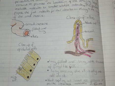
Aug 25, 2021 · The next feature used to increase surface area is called a villus (plural: villi). The villi are small, finger-like projections about a millimeter in length that protrude from the circular folds.
Digestion and absorption
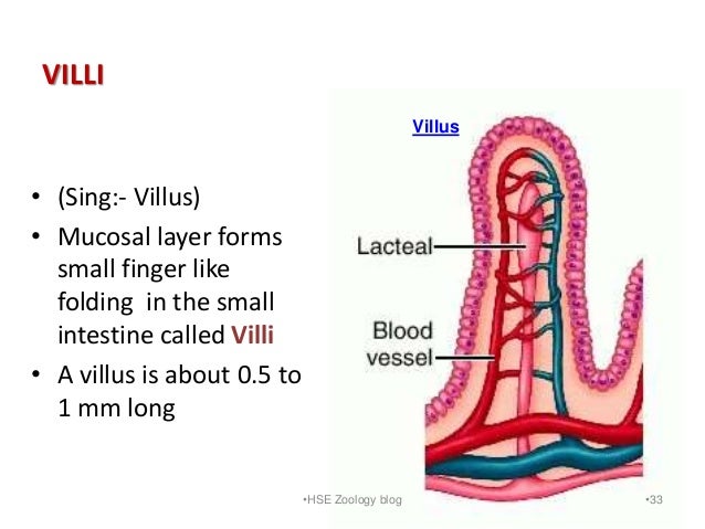
Each villus appears as a finger-like projection from the intestinal lining. They range in length from 0.5 to 1 mm. The villus has several important features: The walls of the villi are one cell thick.

Image from page 123 of "The outlines of anatomy, physiology, and hygiene. Being an edition of The essentials of anatomy, physiology, and hygiene, rev. to conform to the legislation making the effects of alcohol and other narcotics upon the human system a
Ii)label the diagram of the villus below: As shown in the diagram above, each villus contains a capillary bed and a after crossing the epithelium, most of these molecules diffuse into a capillary network inside the villus, and hence into. 6.1.4 draw and label a diagram of the digestive system.

Image from page 102 of "The human body and health : an elementary text-book of essential anatomy, applied physiology and practical hygiene for schools" (1908)
Amniocentesis and chorionic villus sampling had widely been used for the diagnosis, but there is a small risk of miscarriages between 0.5% to 1%. [44] Several other methods have also been developed and are used for the rapid detection of trisomy 21 both during the fetal life and after birth.

Image from page 157 of "A System of midwifery : including the diseases of pregnancy and the puerperal state" (1875)
Draw a schematic diagram of villus in small intestine. Explain how digestive system coordinate with circulatory system. (AS5)

draw-a-schematic-diagram-of-villus-in-small-intestine-789
Microvilli - Diagrams. Microvilli - Diagram . Picture : Microvilli - forming the brush border of intestine. ... Microvilli - It comes from the Greek word mikros which means small and villus, a Latin word which means hair. (3, 5, and 9) Refer to the table below for a brief comparison between cilia and microvilli.
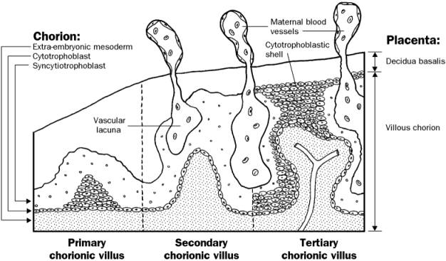
Placenta and Extraembryonic Membranes
1610s, "an illustrative figure giving only the outlines or general scheme of the object;" 1640s in geometry, "a drawing for the purpose of demonstrating the properties of a figure;" from French diagramme, from Latin diagramma "a scale, a musical scale," from Greek diagramma "geometric figure, that which is marked out by lines," from diagraphein "mark out by lines, delineate," from dia "across, through" (see dia-) + graphein "write, mark, draw" (see -graphy). Related: Diagrammatic; diagrammatically. The verb, "to draw or put in the form of a diagram," is by 1822, from the noun. Related: Diagrammed; diagramming.

Image from page 118 of "A treatise on the science and practice of midwifery" (1880)
The main cell type of the duodenal gland is undifferentiated simple columnar cells. These cells multiply, differentiate, and migrate onto the villus, giving rise to the columnar absorbtive cells and goblet cells. They are pushed towards the tip of the villus by succeeding cells, sloughing off into the duodenum's lumen.

Image from page 100 of "Class-book of physiology : for the use of schools and families : comprising the structure and functions of the organs of man, illustrated by comparative reference to those of inferior animals" (1860)
Intestinal structure: (A) diagram of small intestine ... Schematic representation of the crypt /villus axis ... Tales from the crypt : new insights into intestinal stem ...

Image from page 339 of "Biology of the vertebrates : a comparative study of man and his animal allies" (1949)
Outline Stitch. outline stitch diagram. Credit: Alison Gamm. Pull needle up at A. Insert needle back into fabric at B, about 1⁄4 inch from A. Holding floss out of the way, bring needle back up at C and pull floss through so it lies flat against fabric. Pull gently and with equal tautness after each stitch.

Image from page 339 of "Biology of the vertebrates : a comparative study of man and his animal allies" (1949)
Differences Between Chorionic Villus Sampling and Amniocentesis Private Care & Tests During Pregnancy, in London & UK » Antenatal testing genetic testing techniques are an invaluable method of determining the health and safety of an unborn baby, allowing for a diagnosis of conditions like Fragile X and Down's Syndrome weeks before birth.

Image from page 383 of "Kirkes' handbook of physiology" (1907)
Each villus is richly supplied with blood capillaries and lymph vessel or lacteal. Water, mineral salts and other nutrients are directly absorbed through the mucosa. The amino acids and glucose and fructose pass by diffusion from the mucosa into the blood capillaries of the intestine and finally reach the liver through the hepatic portal vein.
Biology Diagrams: Small Intestine
Figure: Diagram of Microvilli. Microvilli form a rather polymorphic class of surface protuberances that are regularly packed in some tissues and loosely positioned in others. Generally, they are shorter and smaller in diameter than cilia. They are commonly about 0.1 µm diameter and range in length from a fraction of a micrometer to about 2 µm.

Image from page 93 of "The essentials of health. A text-book of anatomy, physiology, hygiene, alcohol, and narcotics" (1892)
In fact, it is the longest portion of the digestive system, approximately 20 to 25 feet in length. 1. It is referred to as the "small" intestine because its lumen (opening) is smaller in diameter (at approximately 2.5 centimeters or 0.98 inches) than the large intestine ( colon ). The primary function of the small intestine is to break down ...

Image from page 180 of "Animal biology" (1938)
The diagram below shows the ages at which infants are able to walk unaided. The left end of the bar shows the age at which 25% of infants can walk unaided. The right end of the bar shows the age at which 90% of infants can walk unaided. The vertical line on the bar shows the age at which 50% of infants can walk unaided. 10 111314 1512 Age (months)
![Image from page 346 of "Elements of biology; a practical text-book correlating botany, zoology, and human physiology" ([c1907])](http://live.staticflickr.com/5715/21230952532_ebea6a93f9_c.jpg)
Image from page 346 of "Elements of biology; a practical text-book correlating botany, zoology, and human physiology" ([c1907])
There are about 10-40 villi for every square millimetre of intestinal surface. The centre of each villus has an artery, vein, lacteal, muscle, and connective ...
Toxins | Free Full-Text | Bacterial Heat-Stable ...
... how to draw the villus easily and simply, villus diagram with in 5 ... hd diagram of villus , hd video for villus drawing and structure, ...

Lecture 13: Esophagus & Gastrointestinal Tract
Pinocytosis is a type of endocytosis involved in the transport of particles sized 0.5 µm or less. Process of intake. Phagocytosis involves the formation of pseudopodia (false feet) for the intake of molecules. Pinocytosis intakes the particles via invagination and formation of pockets in the cell membrane.

Image from page 309 of "A civic biology : presented in problems" (c1914)
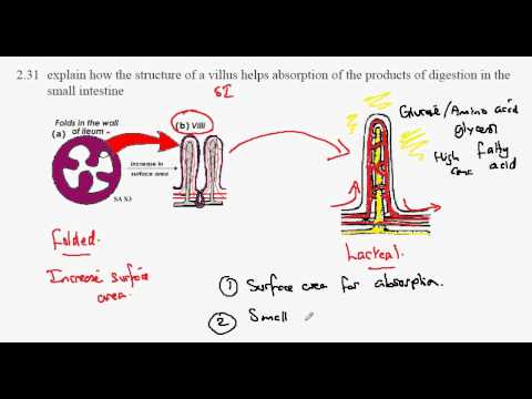
b The diagram shows a cell in the wall of a villus in the small intestine. Some of the processes involved in the absorption of glucose are also shown. - +~~~~~ ~ - ~diffusion of glucose ~ into cell ~ microvfni ..C::::: ~ - - - - - ' What is the importance of the small intestine having villi? (1)

Image from page 391 of "The anatomy of the human body" (1844)
The inner wall of the small intestine is covered by numerous folds of mucous membrane called plicae circulares. The surface of these folds contains tiny ...
2.31 Villi structure and fuction - YouTube
Inside the Villus. Virtally all nurtients, including all amino acids and sugars, enter the body across the epithelium covering small intestinal villi. As shown in the diagram above, each villus contains a capillary bed and a blunt-ended lymphatic vessel referred to as the "central lacteal".
7 _Human nutrition - BIOLOGY4IGCSE
Intestinal villus: An image of a simplified structure of the villus. The thin surface layer appear above the capillaries that are connected to a blood vessel. The lacteal is surrounded by the capillaries. Digested nutrients pass into the blood vessels in the wall of the intestine through a process of diffusion. The inner wall, or mucosa, of the ...

Coeliac Disease Normal Villi And Villous Atrophy Stock ...
1918 (Venn's diagram is from 1904), named for English logician John Venn (1834-1923) of Cambridge, who explained them in the book "Symbolic Logic" (1881).

Image from page 479 of "First course in biology" (1908)
The villus of the jejunum fold lines with the simple columnar epithelium. Within each villus of the jejunum, you will find the lamina propria. At the bottom of the villi, there presence intestinal glands. ... In the labeled diagram, I tried to show you all these above-mentioned histological features from the jejunum.

Image from page 147 of "The development of the human body; a manual of human embryology" (1902)
The diagram illustrates an absorptive structure in the duodenum. What does "A" represent? a. microvilli b. epithelial cell c. capillary d. lacteal e. villus Nutrition includes the study of a. the nutrients in foods b. the body's utilization of nutrients c. how nutrients play a role in your body's health d. digestion, absorption, and transportation […]


Comments
Post a Comment