41 vertebrae diagram unlabeled
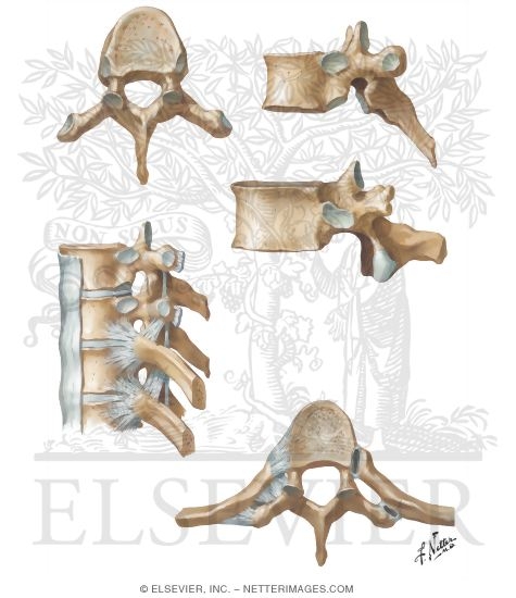
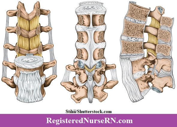
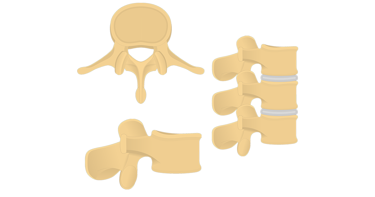
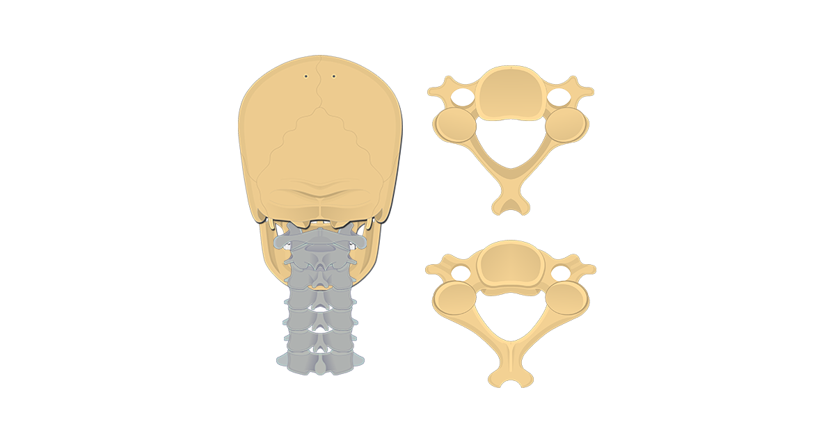
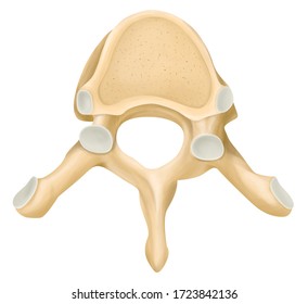
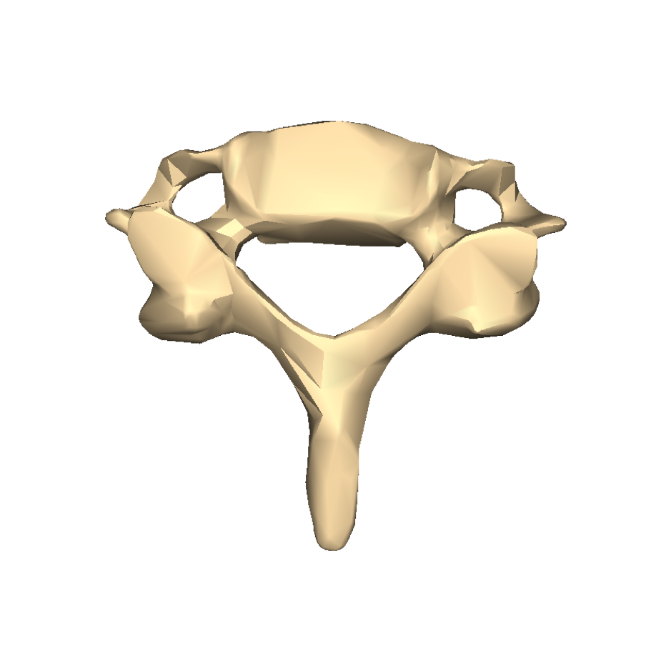
Distinguishing features between the cervical, thoracic, and lumbar vertebrae include: a forked spinous process on vertebrae C2-6, transverse foramen and a large vertebral foramen for the cervical vertebrae. Thoracic vertebrae have relatively pointed and downwards-facing spinous processes, a heart-shaped, body, and transverse costal facets on T1-10. Vertebral Column 17 Vertebrae ... Q. Superior orbital fissure R. Temporal S. Zygomatic T. Zygomatic process of temporal 13 | S k e l e t a l S y s t e m . Artificial Exploded Human Skull (Somso QS 9) Somso Model QS 9 . All individual bones on a plastic base, corresponding
Oct 21, 2021 - Explore Carol Potharaju's board "unlabeled Anatomy" on Pinterest. See more ideas about anatomy, anatomy and physiology, human anatomy and physiology.
Vertebrae diagram unlabeled
diagram labeled unlabeled and, spine anatomy mayfield brain amp spine cincinnati, spine anatomy worksheet northcentral technical college, parts of the spine diagram downloaddescargar com, vertebra wikipedia, the human spine vertebrae numbers anatomy cervical diagram, blank bone diagram rightarrow Learn the vertebral column anatomy of the spine or backbone, including the vertebrae or bones of the cervical, thoracic, lumbar, and sacral spine. Vertebrae by definition function to protect the spinal cord and are located around the cord. Learn the anatomy of the spinal column and each vertebra using labeled diagrams and charts. Start studying Unlabeled Vertebra. Learn vocabulary, terms, and more with flashcards, games, and other study tools.
Vertebrae diagram unlabeled. Hello there, At page below we deliver you various amazing images that we collected in case you need them, for today we are focused related with Vertebrae Labeling Worksheet. While we talk about Vertebrae Labeling Worksheet, scroll down to see some similar photos to inform you more. skeletal system labeling worksheets, sensory and motor neurons spinal cord anatomy and unlabeled vertebral column ... Skeletal system diagrams are illustrations of the human skeleton, used mostly for educational purposes or in presentations. Skeletal diagrams are tools used by students to learn and study all 206 bones (this number can vary from person to person) of the human body. There are four main categories of bones: long, short, irregular, and flat. Check out and save these unlabeled eye diagrams for any evalutional purpose. Eye - The two eyes are located towards the front. This shows you all of the structures youve just learned about in the video labeled on one diagram. Ear diagram not labeled. Cheliped - One of two big claws used for defense and food handling. Gray's Anatomy: The Vertebral Column - The 1917 Gray's Anatomy is available via the Bartleby project. It is available with full colour diagrams. The initial version of this article was copied and pasted from the 1917 Gray's anatomy, which is in the public domain.
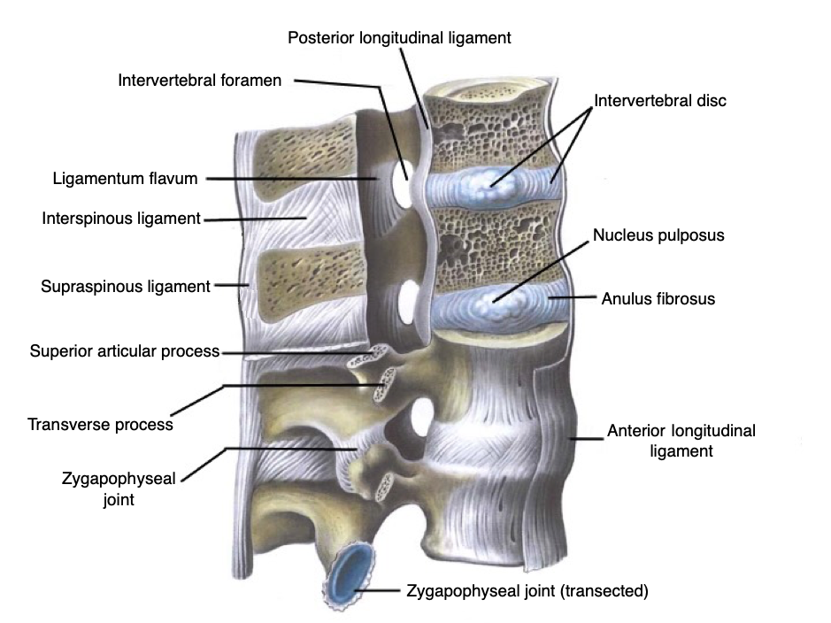
The vertebral foramen is a large, triangular opening in the center of the vertebra that provides space for the spinal cord, cauda equina, and meninges as they pass through the lower back. Extending from the vertebral arch are several bony processes that are involved in muscle attachment and movement of the lower back. The page you tried was not found. You may have used an outdated link or may have typed the address (URL) incorrectly. You might find what you're looking for in one of these areas: · For Technical Support, visit the MH Education Support site at www.mhhe.com/support A basic human skeleton is studied in schools with a simple diagram. It is also studied in art schools, while in-depth study of the skeleton is done in the medical field. This article explains the bone structure of the human body, using a labeled skeletal system diagram and a simple technique to memorize the names of all the bones. A free website study guide review that uses interactive animations to help you learn online about anatomy and physiology, human anatomy, and the human body systems. Start Learning now!
Oct 28, 2021 · Learn anatomy of the spine: Diagrams and interactive vertebrae quizzes. The vertebral column, also known as the spine, is probably the most weird and wonderful looking structure of the human anatomy. It is the home and protector of the spinal cord, but also supports the weight of the upper body, maintains posture and facilitates movement. Nov 28, 2016 - unlabeled diagrams of the human body | Jennifer blog Advanced Search · About Us · Books · Sign In · Register · Artists · John A. Craig Each scapula is a triangular bone and the three edges are known as the superior border, the. Distinguishing features between the cervical, thoracic, and lumbar vertebrae include: a forked spinous process on vertebrae C2-6, transverse foramen and a large vertebral foramen for the cervical vertebrae. Thoracic vertebrae have relatively pointed and ...
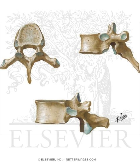
Lumbar Vertebrae Anatomy
To experience full interactivity, please enable Javascript in your browser
Lumbar Vertebrae and Intervertebral Disc Spine: Osteology
Unlabeled Vertebral Column Diagram via. Cervical Vertebrae Blank Diagram via. Femur Bone Diagram Unlabeled via. Human Body Muscles via. Human Body Muscles via. This website is consists of people that are very appreciate creativity from every one, with no exception. That is the reason we always keep the original photos without any editing ...

Lab Exam 2, Thoracic Vertebra Diagram | Quizlet
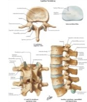
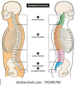
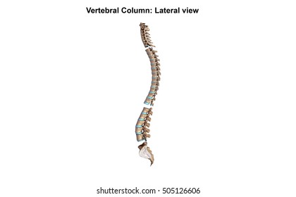
The vertebral column is also known as the spinal column or spine (Figure 1). It consists of a sequence of vertebrae (singular = vertebra), each of which is separated and united by an intervertebral disc. Together, the vertebrae and intervertebral discs form the vertebral column.

12 Skeleton ideas | anatomy bones, anatomy and physiology ...
Find Lumbar spine anatomy diagram stock images in HD and millions of other royalty-free stock photos, illustrations and vectors in the Shutterstock collection. Thousands of new, high-quality pictures added every day.

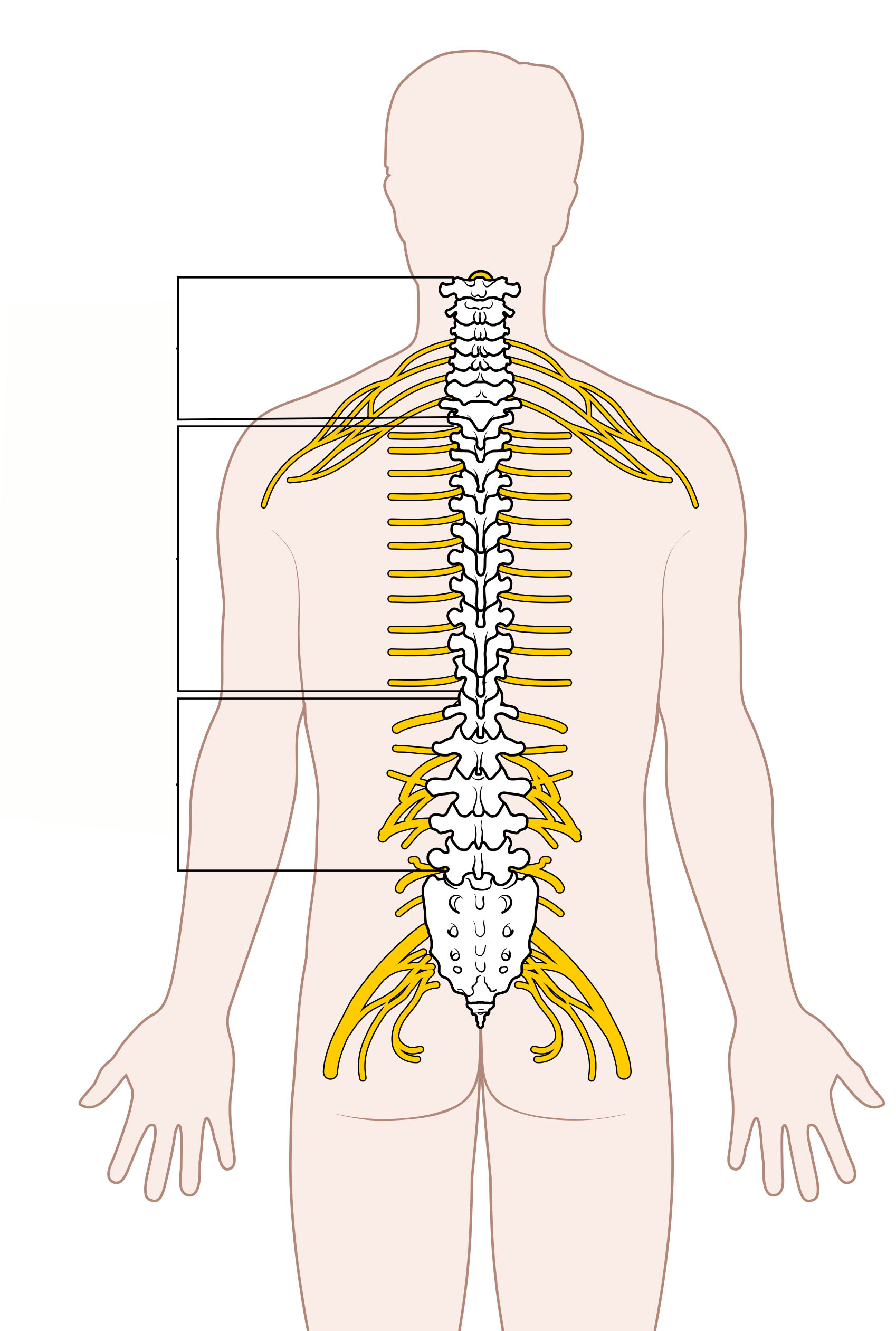
Relation of Spinal Nerve Roots to Vertebrae The Spine
The bones shown in the chest and hip region in the labeled human skeleton diagram are the ribs, vertebrae, pelvis, OS coxae, sacrum and coccyx. Total there are 12 pairs of ribs, as you can see in the diagram. The last pair of the ribs, which is at the bottom of the rib, are called floating ribs, as they are not attached to the sternum.

Cervical Vertebrae Diagram Diagram | Quizlet
September 15, 2014 - Cervical Vertebrae Print Page_ Unlabeled Diagram (Image) and Text - Free download as PDF File (.pdf), Text File (.txt) or read online for free. anatomy
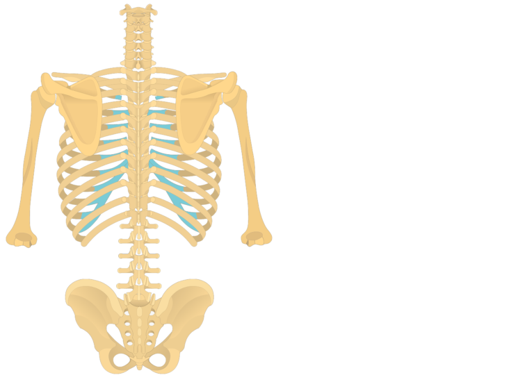
Thoracic Vertebrae and Rib Attachments
Find stockbilleder af Vertebral Column Human Body Anatomy Infograpic i HD og ... Human Heart Muscle Gross Anatomy vector diagram unlabeled outside view with ...

Sacrum and Coccyx Vertebrae Print Page: Unlabeled Diagram and ...
April 20, 2020 - The twelve thoracic vertebrae make up the middle portion of the vertebral column. Review the anatomical characteristics of the vertebrae and test yourself.
Vertebral Column: Anatomy, vertebrae, joints & ligaments | Kenhub
Basic anatomy and parts of the femur. The femur is a long bone found in the lower extremity. It serves as the attachment site for several muscles of the hip and leg, allowing it to withhold pressure from multiple angles. At the proximal end of the femur, it connects with the acetabulum of the pelvis to form the acetabulofemoral joint (aka: the ...

Vertebrae (Photos) - Gross Anatomy Flashcards | Draw it to ...
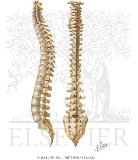
This Illustration was published in ; Atlas of Human Anatomy - 3E · Frank H. Netter · Back and Spinal Cord Page:147 ; Atlas of Human Anatomy - 4th Edition · Frank H.

Lumbar vertebra, illustration - Stock Image - C039/2050 ...
Quiz - Cervical Vertebrae Anatomy (C3 to C7) Learn anatomy faster and remember everything you learn. Start Now. Quiz - Cervical Vertebrae Anatomy (C3 to C7) Start Quiz Retake Quiz. Want to save time learning vertebrae anatomy? Check out these interactive, spaced repetition-inspired quizzes ...
Lumbar Vertebrae and Intervertebral Disc Spine: Osteology
collapsed thoracic vertebrae diagram: thoracic vertebrae diagram unlabeled: 12 3 4 5. Next. People also search for. Thoracic vertebrae. Vertebrae between the cervical vertebrae and the lumbar vertebrae. In vertebrates, thoracic vertebrae compose the middle segment of the vertebral column, between the cervical vertebrae and the lumbar vertebrae ...
Thoracic Vertebrae
This quiz is incomplete! To play this quiz, please finish editing it. 31 Questions Show answers. Question 1. SURVEY. 30 seconds. Q. Which bone is labeled A. answer choices.

Spinal Anatomy | Vertebral Column
spinal cord unlabeled study sheet Spinal Nerves Anatomy, Nerve Anatomy, . somatic Nervous System | Spinal Cord Cross Section Diagram Spinal Cord. A trivia quiz called Cross Section of Spinal Cord and Vertebrae.

Chapter 7: Cervical Vertebrae Diagram | Quizlet
May 15, 2019 - Vertebrae 3D models ready to view, buy, and download for free.

Anatomy Lab Photographs Vertebrae
Anterior · Posterior · Lateral Left
The Vertebral Column | Bones of the Spine | Geeky Medics
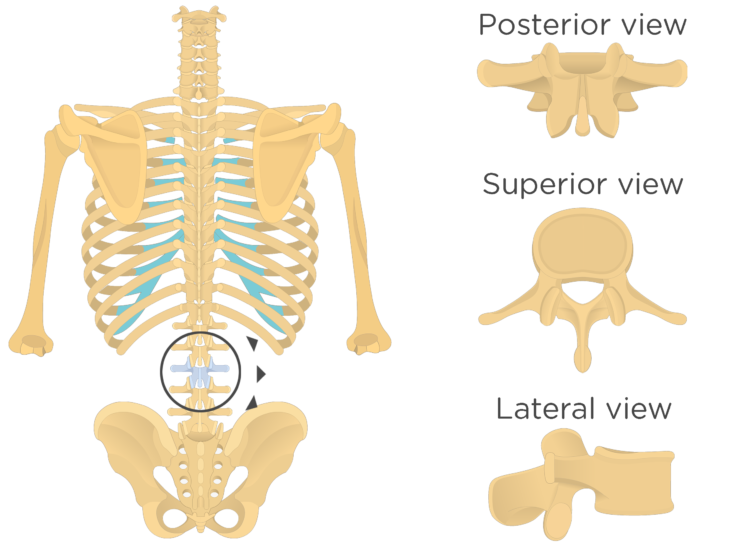
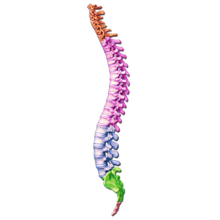
Vertebral Column. Cervical vertebrae (7) Thoracic vertebrae (12) Lumbar vertebrae (5) Sacrum (1) Coccyx (1) Thoracic Cage. Sternum (1) Ribs (24) « Previous (Divisions of the Skeleton) Next (Appendicular Skeleton (126 bones)) ...
Vertebral Labeling Quiz
March 8, 2021 - Here are some blank diagrams I whipped up for drawing in spinal cord pathways. This one shows the whole cord, brainstem, thalamus, and cerebral cortex in coronal section, in cartoon form. It's for drawing in ascending sensory and descending motor pathways, as shown in this office hours sketch.
Vertebral Column Ligaments Quiz
Please Note: You may not embed one of our images on your web page without a link back to our site. If you would like a large, unwatermarked image for your web page or blog, please purchase the appropriate license.

Anatomy Lab Photographs Vertebrae
The vertebral column is also known as the spinal column or spine (Figure 1). It consists of a sequence of vertebrae (singular = vertebra), each of which is separated and united by an intervertebral disc. Together, the vertebrae and intervertebral discs form the vertebral column.

Backbone (Vertebral Column) Labeling Page | Anatomy coloring ...
its unlabeled, so that your practce better. carotid canal coronal suture ethmoid bone external occipital protuberance foramen lacerum foramen magnum foramen
Vertebral Column: The Spine
A posterior view skeletal diagram provides a back view of the human skeleton. The central nervous system lies largely within the axial skeleton the brain being well protected by the cranium and the spinal cord by the vertebral column by means of the bony neural arches the arches of bone that encircle the spinal cord and the intervening ligaments.
Vertebrae cross section Images, Stock Photos & Vectors ...
Printable human skeleton diagram labeled unlabeled and blank. There are four main categories of bones. ... The bones shown in the chest and hip region in the labeled human skeleton diagram are the ribs vertebrae pelvis os coxae sacrum and coccyx. Students fill in the boxes with the names of the bones.

Device for implanting in a human or animal vertebral column ...
Return to Menu. Skeleton. Anterior. Posterior. Lateral Left. Anterior. Lateral Left. Posterior. Vertebral Column Labeling. 1. Select An Answer, Axis(C2) ...
Quiz - Lumbar Vertebrae Anatomy L1 to L5
Just in front of the vertebral column The three short unlabelled arrows indicate that the mesoderm is being subdivided into three columns. Royalty free no fees and download now in the size you need. This diagram depicts human body map of organs with parts and labels. Cranial cavitythe space occupied by the brain enclosed by the skull bones.
Quiz - Cervical Vertebrae Anatomy (C3 to C7)
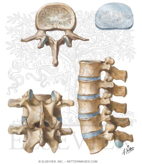
The rib cage is joined to the thoracic vertebrae. At T11 and T12, the ribs do not attach and are so are called "floating ribs." The thoracic spine's range of motion is limited due to the many rib/vertebrae connections and the long spinous processes. Lumbar Vertebrae (L1 - L5) The lumbar vertebrae graduate in size from L1 through L5.
Vertebras Images, Stock Photos & Vectors | Shutterstock
April 21, 2020 - Inferior to the atlas bone (C1) and axis bone (C2) are the remaining five cervical vertebrae (C3-C7). The vertebrae share many anatomical characteristics. Click and start learning now!

✓ Unlabeled Vertebra Cross Section of Human Body Anatomy ...
Brain and Vertebrae Unlabelled Diagrams. School University of New South Wales. Course Title ANAT 1521. Uploaded By emilyyxo. Pages 4. This preview shows page 1 - 4 out of 4 pages. View full document. End of preview.
Vertebral anatomy lateral view Images, Stock Photos & Vectors ...
The third curve of the spine that is composed of 5 vertebrae in the lower back. The vertebral column is bony it is strong yet it is flexible as it comprises a number of smaller bones. Unlabeled vertebral column worksheet. Do you know how many vertebrae are in the vertebral column and where are the joints of Luschka found.
The Vertebral Column | Bones of the Spine | Geeky Medics
20. aug. 2018 ... Unlabeled Vertebra Cross Section of Human Body Anatomy infographic diagram including all parts cord of grey and white matter spinal nerve ...
Skeleton Worksheet - WikiEducator
Please Note: You may not embed one of our images on your web page without a link back to our site. If you would like a large, unwatermarked image for your web page or blog, please purchase the appropriate license.

T III. Thoracic Vertebra, Superior view. on Behance
Start studying Unlabeled Vertebra. Learn vocabulary, terms, and more with flashcards, games, and other study tools.
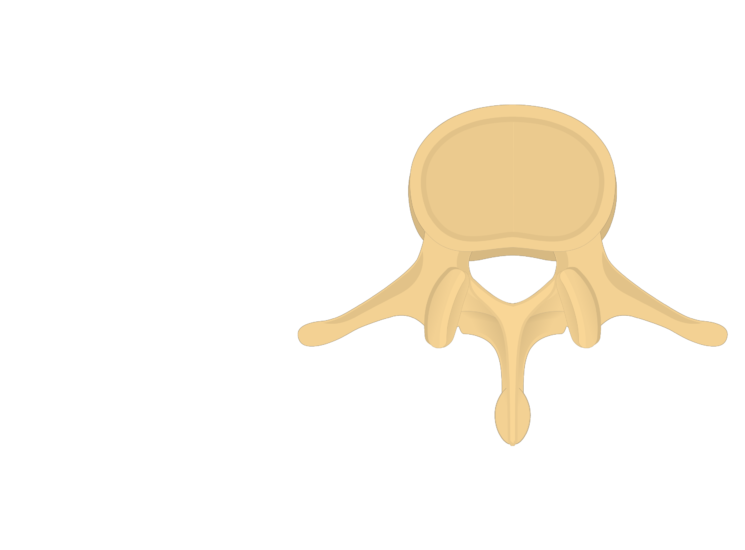
Lumbar Vertebrae Anatomy
Learn the vertebral column anatomy of the spine or backbone, including the vertebrae or bones of the cervical, thoracic, lumbar, and sacral spine. Vertebrae by definition function to protect the spinal cord and are located around the cord. Learn the anatomy of the spinal column and each vertebra using labeled diagrams and charts.
File:Diagram of the Spinal Cord Unlabeled.jpg - Wikimedia Commons
diagram labeled unlabeled and, spine anatomy mayfield brain amp spine cincinnati, spine anatomy worksheet northcentral technical college, parts of the spine diagram downloaddescargar com, vertebra wikipedia, the human spine vertebrae numbers anatomy cervical diagram, blank bone diagram rightarrow

Vector Illustration Of Sacrum And Lumbar Vertebrae In ...
Lumbar Vertebrae Anatomy

Anatomy Lab 2 Practicum (vertebral column) Diagram | Quizlet
Vertebral Column: Anatomy, vertebrae, joints & ligaments | Kenhub

Anatomy Lab Photographs Vertebrae
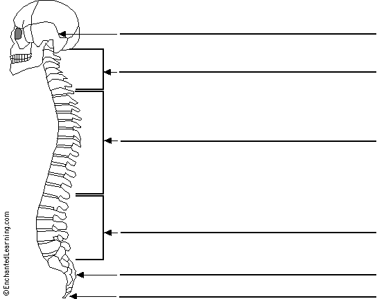
Label the Spine Printout - EnchantedLearning.com

The Skeletal System - Vertebral Column Diagram Diagram | Quizlet


Comments
Post a Comment